At Synnovis, innovation is in our DNA. We’re proud to always be at the cutting edge of modern pathology services, adopting new technologies that enable us to provide an outstanding experience for our service users.
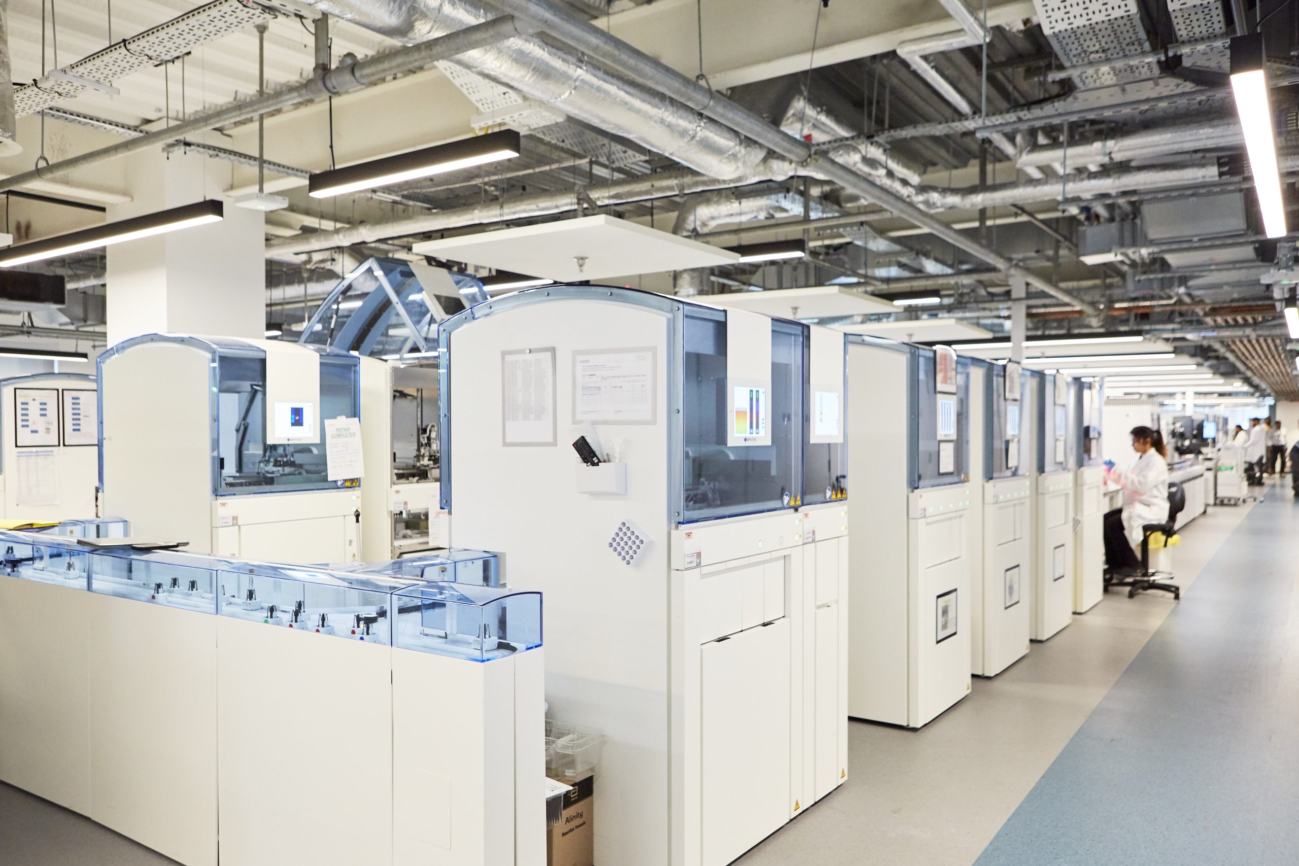
Digital pathology is a key feature of our service offering in south east London, enabling clinicians to review Haematology, Microbiology and Histopathology samples in the form of high-quality digital images from wherever they are when they need them. It reduces the need for on-site review of samples and widens opportunities for seeking a second opinion where needed.
Adopting digital pathology is just one of the ways we’re meeting our commitment to implement modern digital solutions and realise major benefits for clinicians, patients and the wider NHS.
Digital pathology: key benefits
Slides, films and cultures are created, analysed and reported more quickly, facilitating faster diagnoses for thousands of patients, while remote access to samples supports clinical decision-making.
Samples can be processed faster on new automated machinery, with pathologists able to view them from any location immediately after they are scanned, facilitating quicker turnaround times and more flexible remote working arrangements.
Laboratory Information Management System (LIMS) integration will streamline workflows by further reducing manual processes.
All clinicians will be able to view samples during multi-disciplinary meetings and benefit from easier access to second opinions from clinicians anywhere in the world.
We’re also looking ahead to the future, and the opportunities presented by Artificial Intelligence (AI) integration to further streamline the reporting process.
How digital pathology works
Digital Histopathology
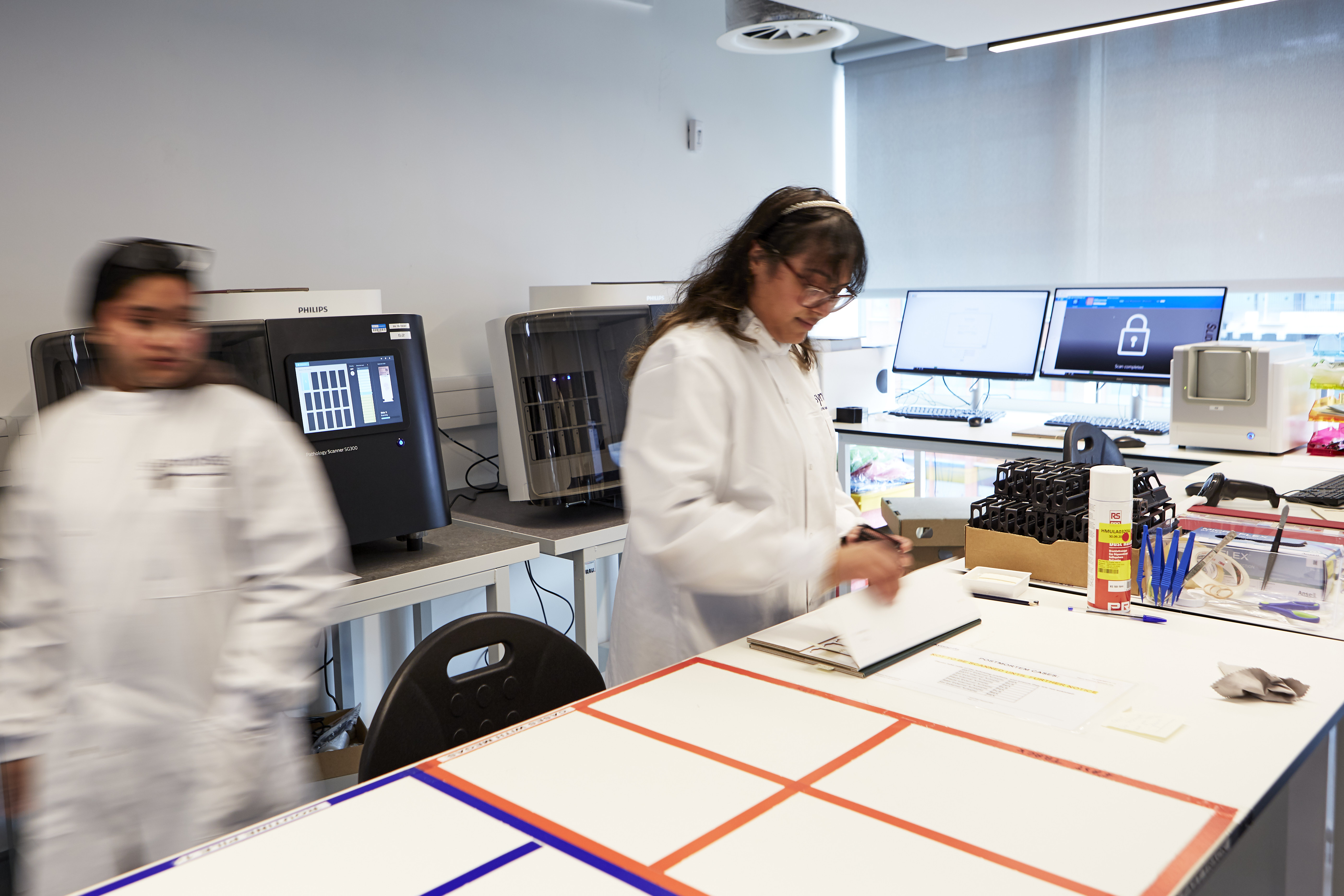
Our hub laboratory is equipped with state-of-the-art Sakura, Roche and Leica machines that fully automate the staining of glass tissue samples slides. All slides produced at the hub are loaded into our Philips scanners to produce high-quality images accessible via the Philips Image Management System (IMS).
Digital slides can be viewed and reported directly to EPIC on high-resolution monitors, with cases available to any clinicians with access to the system, wherever they are.
Digital Haematology
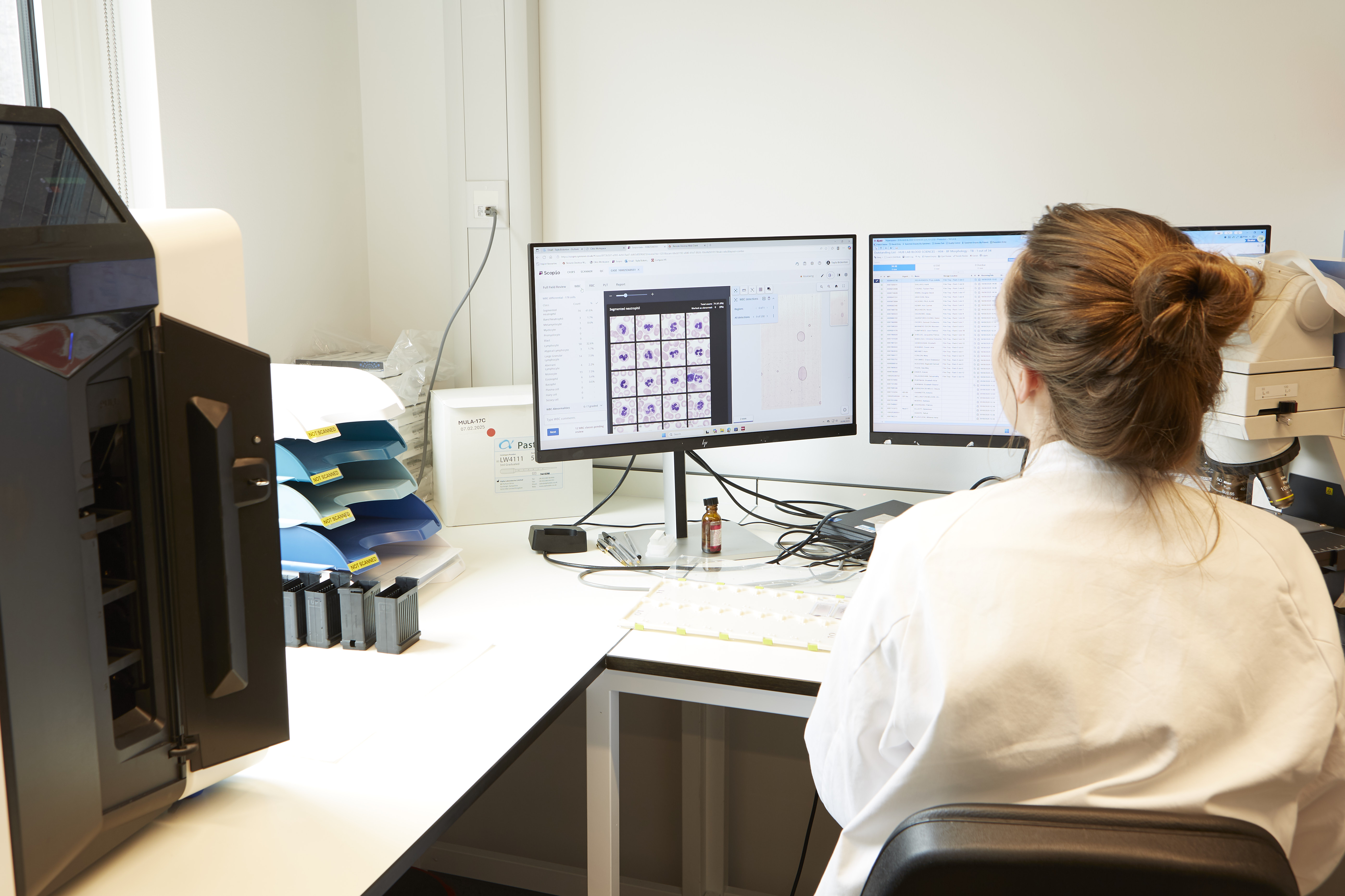
Digital haematology utilises state-of-the-art Scopio scanners and high-resolution monitors installed in our laboratories. They scan blood films to create detailed digital images of samples that can be viewed and reported directly to EPIC via the Scopio app.
This process makes it possible to view whole slides at 100x resolution, with clinicians also able to zoom in to view sharp individual cellular and subcellular detail at 100x magnification.
Our use of digital haematology is ever evolving, with plans to incorporate AI support in the future.
Digital Microbiology
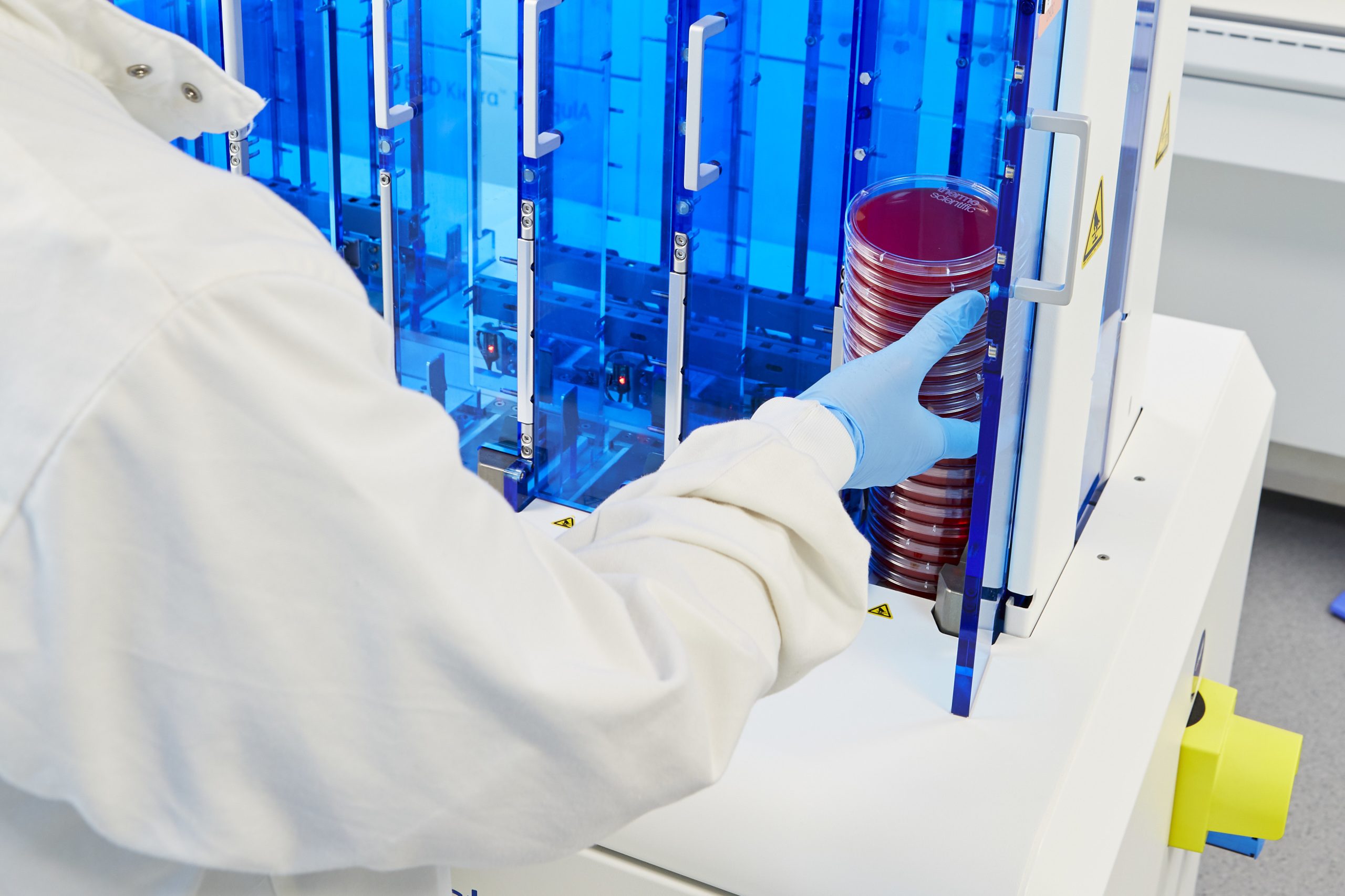
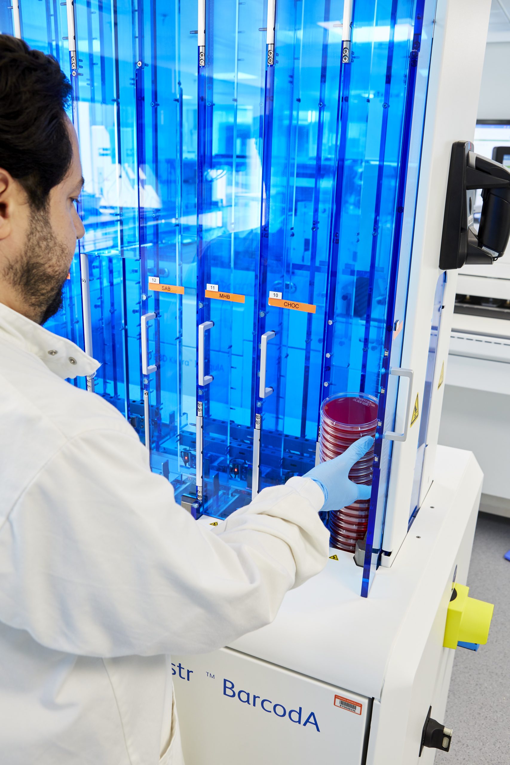
Cultures are still grown in plates, but instead of manual inspection, detailed images of the plates are captured by the scanners in our laboratories. The system uses these images to determine when organisms have grown so they can be reviewed, and further tests can be ordered by scientists and clinicians.